Carbohydrates are digested with juice. All about medicine. Daily intake of carbohydrates
Let's first understand why carbohydrates are needed at all.
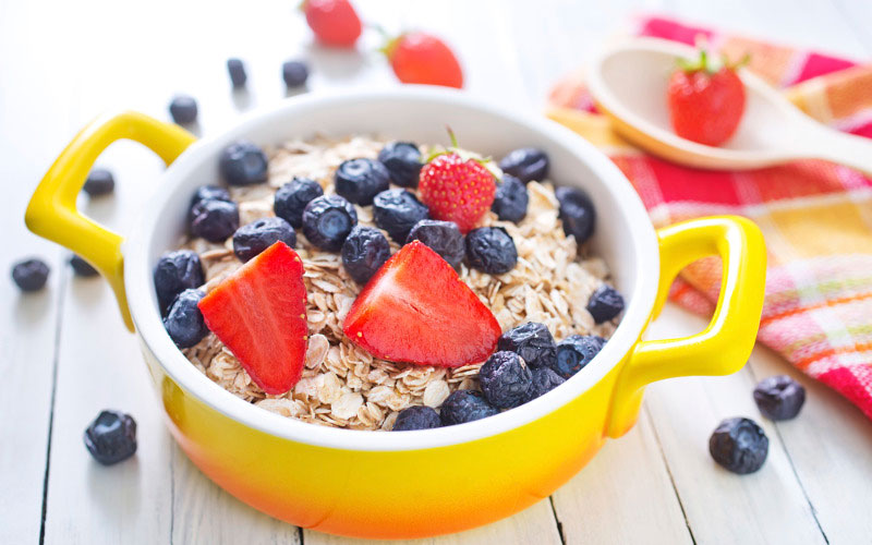
As you know, carbohydrates provide our body with energy, which we spend on all the basic processes: heating the body, physical activity, mental activity. Why, then, nutritionists do not allow us to eat buns or delicious chips? After all, these are also carbohydrates, and they, according to the same nutritionists, are absolutely necessary for us.
The fact is that carbohydrates, according to the rate of assimilation by the body, are divided into fast and slow.
Fast carbohydrates (or simple) - These are those that are absorbed literally as soon as they enter the stomach.
The processing of carbohydrates is responsible for the insulin produced by the pancreas. When we eat a certain amount of sugar, for example, it immediately begins its work: they quickly capture the entire batch of carbohydrates and process it into energy.
And if we did not eat sugar, but say, a donut? Then, along with sugar, a certain (rather impressive) amount of fat entered the body. In this case, insulin removes carbohydrates, saturating the body with energy, and fat process it "laziness". And the truth is, why should he pick his fats if our brain gives a signal that he has received enough fuel.
But insulin is also responsible for the exchange of fats, therefore, without thinking twice, he transfers all the fat to the storage tanks that we see in the mirror or with his own eyes, trying again to fasten the skirt.
And what do we do, do not eat carbohydrates at all? It turns out, no, you need to consume carbohydrates, but it is better to choose slow carbohydrates.
Slow (or complex) carbohydrates- These are those that are absorbed by the body gradually, they secrete sugar slowly, which means that the brain will not give a signal about hunger for longer.

It is this phenomenon that we encounter when we go on a diet. Wrong carbohydrates make us suffer from hunger. The fast ones are instantly absorbed, and the excess is carried off to the pantries. And only the slow ones are able to maintain a feeling of satiety for a long time.
You might have a slightly misconception that fast carbohydrates are sugar. No, there is one more important nuance.
Fast carbohydrates are characterized not only by fast digestibility, but also by the ability to quickly and for a short period of time raise the level of glucose in the blood, which then also drops rapidly.
Slow carbohydrates have a different quality, they do not raise glucose levels so fast and are able to maintain it at the proper level for quite a long time, not allowing the brain to demand more food.
Based on the speed with which carbohydrates are absorbed from certain foods, a new unit was derived - the glycemic index. The higher it is, the faster the carbohydrate.
And the top fast carbohydrates top is not sugar, as you might have guessed. Before him go baked potatoes, corn flakes, honey and other products, including even carrots. What is the reason?
Apparently in starch. You have probably heard the expression “starchy vegetables and fruits”, these include bananas, beets, carrots and much more. So, all this is also fast carbohydrates.
So, choosing a diet for diet food, we need to focus not so much on calories as on the value of the glycemic index of products. Vegetables and fruits, similar in calorie content, can vary significantly in the content of fast and slow carbohydrates. This means that carrot salad will linger in the stomach for a shorter period than, say, fruit from fruits with a low glycemic index.
If you do not go into too much detail, you can highlight the main features of most foods with fast carbohydrates.

First of all, of course, sweet foods . Sugar, although not the first place it takes, but still absorbed quickly enough. If the product tastes sweet, it is sure to have a lot of simple carbohydrates.
Then you should pay attention to, say, product consistency . A hard fruit or vegetable most often contains more slow carbohydrates, and a soft one contains more fast carbohydrates. Although the same carrot in this regard is more harmful than bananas.
The next moment is canned and processed foods. Glucose is often used for preservation, which means that the glycemic index of such food will be higher. The same can be said about fast food products. The amount of simple carbohydrates that you eat with crackers and chips does not compare with what you eat with a plate of fried potatoes with a slice of brown bread.
Of course, by these signs it is good to determine the harmfulness of finished products or food, the calorie content and sweetness of which are not in doubt.
If you are going to go on a diet based on the distinction between fast and slow carbohydrates, then it is better to focus on special tables where the glycemic index of products is indicated. This is in the diet of separate nutrition and others similar to this method.
If you already have experience using this method, we will be glad to hear your recommendations.
fast carbohydrates slow carbohydrates fast and slow carbohydrates
Refined sugar is the sweetest poison
Why is sugar toxic to the body?
In 1957, Dr. William Martin gave the following definition of toxic food: "This is a substance that does not digest or accumulate in the body, which leads or can lead to the development of diseases." Martin defined refined sugar as a toxin, as it is deprived of its vitality, vitamins and minerals. All that remains in such sugar is refined carbohydrates.
Nature supplies vitamins and minerals to each plant in sufficient quantities so that when the plant is used for food, the body can process them. However, all useful substances are lost during the industrial processing of plants. Natural minerals found in sugar beets or cane sugar are not found in refined sugar made from them.
Sugar dries and leaches the body, removing valuable vitamins and minerals from it. These substances are consumed due to sugar, which extracts these substances for their own digestion, further detoxification of the body from the products of sugar digestion and their removal from the body.
Sugar consumed every day gives constant acidity, and more and more minerals need to be delivered from distant parts of the body to the digestive system to restore balance. As a result, a lot of calcium is removed from the bones and teeth, which leads to their destruction, and in general, to weakening the body.
Increased amounts of sugar ultimately affect every organ in the body. Initially, it is deposited in the liver in the form of glycogen. If sugar is consumed every day, the liver will begin to expand. When it reaches its maximum volume, excess glycogen from the liver will enter the blood in the form of fatty acids. They are spread throughout all parts of the body and are deposited in its least active areas: in the abdomen, sciatica, chest, and thighs.
When these relatively safe places are full, fatty acids go to more active organs, such as the heart and kidneys. They begin to slow down their work, their tissues degrade and turn into fat. The whole body begins to suffer from decreased heart and kidney function, which leads to high blood pressure.
The circulatory and lymphatic systems work worse. The body's immune stamina decreases with respect to bacteria, heat, cold, etc.
Fredrik Bunting, one of those who discovered insulin after visiting Panama in 1929, found interesting results by observing workers on sugar plantations. For those who eat refined sugar, diabetes would be widespread. For those who had barely processed sugarcane, this was not observed.
Refined Sugar and Food
At one time, sugar was advertised as a substance that can give a lot of energy. Later, when it became clear that excess calories from sugar did not lead to anything good, they began to advertise sugar as a chemically pure product. As if the words “chemically pure” automatically mean “healthy”. Also often in product advertisements or on packages instead of refined sugar they write - carbohydrates.
In chemistry, sugars are classified as carbohydrates, i.e. compounds consisting of carbon and hydrogen. However, when the word carbohydrates is used in the labeling of food products, without deciphering which particular compounds are in question, this is often misleading.
That is, people often do not know what exactly is part of these carbohydrates in this product. What kind of sugar? There are several types of sugars.
Glucose - sugar, which is found in fruits and vegetables. Glucose is the most important substance involved in the metabolism of all plants and animals. In the human body, glucose also plays a very important role.
Fructose - sugar found in fruits.
Lactose - milk sugar.
Sucrose - Refined sugar (which we are talking about), industrially produced sugar from beets or sugarcane.
If glucose is the most important element for the metabolism in our body, then sucrose is something new and unnatural for the body.
Eating refined sugar and white flour instead of whole flour and natural sugars from fruits and vegetables, we change the balance of metabolism in the body. One spoonful of sugar in coffee after a sandwich is enough to turn your stomach into a fermenter. One can of fizzy drink with a hamburger turns your stomach into a distillery.
How is sugar digested?
Sugars are not digested in the mouth (like grain) or in the stomach (like meat). When eaten separately, they quickly go from the stomach to the small intestine. When sugar is taken with other foods (for example, bread and meat, as in a sandwich), they linger in the stomach for a while. While the stomach is working on the processing of animal proteins and the processing of refined starch, sugar guarantees fast acid fermentation in the stomach, which is completely useless.
When we consume complex sugars, eating fruits or honey, they break down when digested into simple monosaccharides - important substances for our health.
When we consume refined sugar along with other starch-rich foods, a fermentation process starts in the stomach and results in carbon dioxide, acetic acid, alcohol and water. In addition to water, all other products of this process are harmful.
When we eat separately protein-rich foods, they break down in the stomach into amino acids, which are very necessary in metabolism. But if we eat proteins with sugar, ptomains and leukomains are formed in the stomach, harmful substances that are not needed for metabolism.
Read also:
Carbohydrates are the main component of the human diet, because they consume about 4 times more than fats and proteins. They perform many diverse functions in the body, but the main one is energy.
The average need for carbohydrates is 350-500 g / day. Carbohydrates are necessary for the biosynthesis of nucleic acids, interchangeable amino acids as an integral structural part of cells. They have regulatory, protective and plastic significance.
By nutritional value, carbohydrates are divided into digestible and non-digestible. Digestible carbohydrates are digested and metabolized in the human body. These include glucose, fructose, sucrose, lactose, maltose, α-glucan polysaccharides-starch, dextrins and glycogen.
Non-digestible carbohydrates are not broken down by enzymes secreted in the human digestive tract. Indigestible carbohydrates include raffinous oligosaccharides and non-α-glucan polysaccharides - cellulose, hemicellulose, pectin, lignin, gums and mucus.
Digestible Carbohydrates.Aldoses (glucose, galactose, mannose, xylose), as well as ketose (fructose) have the highest nutritional value.
How fast are fast carbohydrates and why are slow carbs so slow? Carve Myths About Carbohydrates!
Consumption of glucose and fructose, the two most common monosaccharides in nature, reaches 20% of the total carbohydrate intake. From the intestines, carbohydrates are absorbed into the blood only in the form of glucose and fructose.
The main food disaccharides in human nutrition are sucrose and lactose.
Sugar, the main component of which is sucrose, plays the role of an energy carrier in the body.
The most frequent and serious consequences of excessive consumption of refined sugar are metabolic disorders, primarily carbohydrate metabolism.
Lactose is the most important carbohydrate during breastfeeding and during artificial feeding of infants. The main sources of lactose in foods are milk (4.8-5.2%), cream (3.7%), sour cream and kefir (3.1-3.6%).
Among the polysaccharides of plant products, starch is of the greatest importance in human nutrition. The absorption of starch requires significantly more time than the absorption of sugar. The final product of the breakdown of starch - glucose - enters the blood slowly, its concentration is maintained at the same level. Most starch is found in bakeries (40-73%), seeds of legumes (40-45%) and potatoes (15%)
In animal products, there is a relatively small amount of another digestible polysaccharide that is close in chemical structure to starch, glycogen (in the liver 2-10%, in muscle tissue 0.3-1.0%)
With a lack of carbohydrates in the body, weakness, dizziness, headache, hunger, drowsiness, sweating, trembling in the hands appear.
Non-digestible carbohydrates. The main non-digestible carbohydrates are the so-called “dietary fiber” - a mixture of various structural polysaccharides of plant cells — cellulose, hemicellulose and pectin, lignin and non-structural polysaccharides. Naturally occurring foods, gums, mucus and polysaccharides used as food additives.
Cellulose is the main structural component of the membrane of a plant cell. Its main physiological effect is the ability to bind water (up to 0.4 g of water per 1 g of fiber).
Hemicelluloses are cell wall polysaccharides consisting of glucose and hexose polymers. They are also able to retain water and bind cations.
Pectin substances - glycanogalacturonans - the main component of plants and algae. One of the most important properties of pectin substances is its complexing ability, based on the interaction of pectin molecules with ions of heavy metals and radionuclides. This gives reason to recommend pectin for inclusion in the diet of people in an environment contaminated with radio nuclides and having contact with heavy metals . The prophylactic norm of pectin, approved by the WHO, is 2-4g per day.
Lignins are carbohydrate-free substances of the cell membrane, consisting of polymers of aromatic alcohols. Lignins in the human body are capable of binding bile salts and other organic substances, as well as slowing down or disrupting the absorption of nutrients in the colon.
Gums are complex unstructured polysaccharides, they are soluble in water, have viscosity, contain glucuronic and galacturonic acids, are able to participate in the binding of trace elements with even valency.
Thus, dietary fiber is one of the components of the comprehensive prevention of disorders of fat metabolism, atherosclerosis, diabetes mellitus, and gallstone disease. In recent years, evidence has appeared that a lack of dietary fiber causes the development of urolithiasis, gastric and duodenal ulcers, gout, caries, and even varicose veins.
The daily norm of dietary fiber for an adult is 25-30 g.
The main source of dietary fiber is cereal products, fruits, nuts and vegetables.
Dietary fiber affects the function of the large intestine. They stimulate peristalsis, increase the secretion of bile.
At the same time, excess fiber intake is more harmful than beneficial. It can lead to incomplete digestion of food, impaired intestinal absorption of macro- and microelements, as well as fat-soluble vitamins. Excessive intake of dietary fiber causes diarrhea, discomfort from excessive gas formation in the intestines, and abdominal pain.
Only monosaccharides are absorbed in the intestines: glucose, galactose, fructose. Therefore, the oligo- and polysaccharides that enter the body with food must be hydrolyzed by enzyme systems to form monosaccharides. In fig. 5.11 schematically depicts the localization of enzymatic systems involved in the digestion of carbohydrates, which begins in the oral cavity with the action of oral -amylase and then continues in different parts of the intestine using pancreatic -amylase, sucrose-isomaltase, glycoamylase, -glycosidase (lactase) trehalase complexes.
Fig. 5.11. The scheme of localization of enzyme systems for the digestion of carbohydrates
5.2.1. Digestion of carbohydrates using oral and pancreatic amylase ( -1,4-glycosidases).Food polysaccharides, namely starch (consists of a linear amylose polysaccharide, in which glucose residues are linked with 1,4-1,4-glycoside bonds, and amylopectin, a branched polysaccharide, where -1,6-glycoside bonds are also found) begin to hydrolyze already in the oral cavity after wetting with saliva containing the hydrolytic enzyme -amylase (-1,4-glycosidase) (KF 3.2.1.1), which breaks down 1,4-glycosidic bonds in starch, but not acting on 1,6-glycosidic bonds.


In addition, the contact time of the enzyme with starch in the oral cavity is short, so the starch is partially digested, forming large fragments dextrins and a little maltose disaccharide. Disaccharides are not hydrolyzed by saliva amylase.
When saliva amylase enters the stomach in an acidic environment, the digestion process can only occur inside the food coma, where the amylase activity can remain for some time until the pH in the whole piece is acidic. In the gastric juice there are no enzymes that break down carbohydrates, only slight acid hydrolysis of glycosidic bonds is possible.
The main place for the hydrolysis of oligo- and polysaccharides is the small intestine, in which certain glycosidases are secreted in different departments.
In the duodenum, the contents of the stomach are neutralized by pancreatic secretions containing HCO 3 bicarbonates and having a pH of 7.5–8.0. Pancreatic secretion reveals pancreatic amylase, which hydrolyzes -1,4-glycosidic bonds in starch and dextrins with the formation of maltose disaccharides (in this carbohydrate two glucose residues are linked by a 1,4-1,4-glycosidic bond) and isomaltoses (there are two glucose residue located at the branching sites in the starch molecule and linked by 1,6-1,6-glycosidic bonds). Oligosaccharides are also formed with 810 glucose residues linked by both -1,4-glycosidic and -1,6-glycosidic bonds.
Both amylases are endoglycosidases. Pancreatic amylase also does not hydrolyze -1,6-glycosidic bonds in starch and -1,4-glycosidic bonds, by which glucose residues are connected in a cellulose molecule.

Cellulose passes through the intestines unchanged and serves as a ballast substance, giving food volume and promoting the digestion process. In the large intestine, under the influence of bacterial microflora, cellulose can partially hydrolyze with the formation of alcohols, organic acids and CO 2, which can act as stimulants of intestinal motility.
Maltose, isomaltose, and triozosaccharides formed in the upper intestine are then hydrolyzed in the small intestine by specific glycosidases. Disaccharides of food, sucrose and lactose, are also hydrolyzed by specific small intestine disaccharidases.
In the intestinal lumen, the activity of oligo- and disaccharidases is low, but most of the enzymes are associated with the surface of epithelial cells, which are located in the intestine on finger-like outgrowths villi and themselves, in turn, are covered with microvilli, all these cells form a brush border that increases the contact surface of hydrolytic enzymes with their substrates.
The cleaving glycosidic bonds in disaccharides, enzymes (disaccharidases) are grouped into enzyme complexes located on the outer surface of the cytoplasmic membrane of enterocytes: sucrose-isomaltase, glycoamylase, -glycosidase.
5.2.2. Sugara-isomaltase complex. This complex consists of two polypeptide chains and attaches to the surface of the enterocyte using a transmembrane hydrophobic domain located at the N-terminal portion of the polypeptide. The sucrose-isomaltase complex (K.F. 3.2.1.48 and 3.2.1.10) cleaves -1,2- and -1,6-glycosidic bonds in sucrose and isomaltose.


Both enzymes of the complex are also able to hydrolyze -1,4-glycosidic bonds in maltose and maltotriosis (a trisaccharide containing three glucose residues and formed during the hydrolysis of starch).

Although the complex has a rather high maltase activity, hydrolyzing 80% of the maltose formed during the digestion of oligo- and polysaccharides, its main specificity is nevertheless hydrolysis of sucrose and isomaltose, the rate of hydrolysis of glycosidic bonds in which is greater than the rate of hydrolysis of bonds in maltose and maltotriosis. In this case, the sucrose subunit is the only intestinal enzyme that hydrolyzes sucrose. The complex is localized mainly in the jejunum, in the proximal and distal parts of the intestine, the content of the sucrose-isomaltase complex is negligible.
5.2.3. Glycoamylase complex. This complex (KF 3.2.1.3 and 3.2.1.20) hydrolyzes -1,4-glycosidic bonds between glucose residues in oligosaccharides. The amino acid sequence of the glycoamylase complex has 60% homology with the sequence of the sucrose-isomaltase complex. Both complexes belong to the family of 31 glycosyl hydrolases. Being an exoglycosidase, the enzyme acts from the reducing end and can also break down maltose, acting as a maltase in this reaction (the glycoamylase complex hydrolyzes the remaining 20% \u200b\u200bof the maltose oligo- and polysaccharides formed during digestion). The complex consists of two catalytic subunits that have small differences in substrate specificity. The complex is most active in the lower parts of the small intestine.
5.2.4. -Glycosidase complex (lactase). This enzyme complex hydrolyzes -1,4-glycosidic bonds between galactose and glucose in lactose.

The glycoprotein is associated with the brush border and is unevenly distributed throughout the small intestine. With age, the activity of lactase decreases: it is maximum in infants, in adults it is less than 10% of the level of activity of the enzyme excreted in children.
5.2.5. Tregalase. This enzyme (K.F. 3.2.1.28) is a glycosidase complex that hydrolyzes bonds between monomers in trehalose, a disaccharide found in fungi and consisting of two glucosyl residues linked by a glycosidic bond between the first anomeric carbon atoms.

Monosaccharides are formed from food carbohydrates as a result of the action of glycosyl hydrolases: in a large amount of glucose, fructose, galactose, to a lesser extent mannose, xylose, arabinose, which are absorbed by the epithelial cells of the jejunum and ileum and are transported through the membranes of these cells using special mechanisms.
5.2.6. Transport of monosaccharides through the membrane of intestinal epithelial cells.The transfer of monosaccharides into the cells of the intestinal mucosa can be carried out by facilitated diffusion and active transport. In the case of active transport, glucose is transported through the membrane along with the Na + ion by one carrier protein, and these substances interact with different parts of this protein (Fig. 5.12). The Na + ion enters the cell according to the concentration gradient, and glucose against the concentration gradient (secondary active transport), therefore, the larger the gradient, the more glucose is transferred to enterocytes. With a decrease in the concentration of Na + in the extracellular fluid, glucose intake decreases. The concentration gradient of Na + underlying the active symport is ensured by the action of Na +, K + -ATPase, which works as a pump pumping Na + out of the cell in exchange for the K + ion. In the same way, by the mechanism of secondary active transport, galactose enters enterocytes.

Fig. 5.12. The entry of monosaccharides into enterocytes. SGLT1 sodium-dependent glucose / galactose transporter in the epithelial cell membrane; Na +, K + -ATPase on the basolateral membrane creates a concentration gradient of sodium and potassium ions necessary for the functioning of SGLT1. GLUT5 transports primarily fructose through the membrane into the cell. GLUT2 on the basolateral membrane transports glucose, galactose and fructose from the cell (according to)
Thanks to active transport, enterocytes can absorb glucose at its low concentration in the intestinal lumen. At a high concentration of glucose, it enters the cells by facilitating diffusion using special carrier proteins (transporters). In the same way, fructose is transferred into the epithelial cells.
Monosaccharides enter blood vessels from enterocytes mainly through facilitated diffusion. Half of the glucose through the capillaries of the villi through the portal vein is transported to the liver, half is delivered by blood to the cells of other tissues.
5.2.7. Transport of glucose from blood to cells. The flow of glucose from the blood into the cells is carried out by facilitated diffusion, i.e., the rate of glucose transport is determined by the gradient of its concentrations on both sides of the membrane. In muscle cells and adipose tissue, facilitated diffusion is regulated by pancreatic hormone insulin. In the absence of insulin, the cell membrane does not contain glucose transporters. The protein carrier (transporter) of glucose from red blood cells (GLUT1), as can be seen from Fig. 5.13, is a transmembrane protein consisting of 492 amino acid residues and having a domain structure. Polar amino acid residues are located on both sides of the membrane, hydrophobic are localized in the membrane, crossing it several times. There is a glucose binding site on the outside of the membrane. Upon glucose binding, the carrier conformation changes, and the monosaccharide binding site is open inside the cell. Glucose passes into the cell, separating from the carrier protein.
5.2.7.1. Glucose transporters: GLUT 1, 2, 3, 4, 5. In all tissues, glucose transporters were found, of which there are several varieties, which were numbered in the order of their detection. Five types of GLUT are described, having a similar primary structure and domain organization.
GLUT 1, located in the brain, placenta, kidneys, large intestine, red blood cells, provides glucose to the brain.
GLUT 2 transfers glucose from the organs that secrete it into the blood: enterocytes, liver, transports into the клетки-cells of the pancreatic islets of Langerhans.
GLUT 3 is found in many tissues, including the brain, placenta, kidneys, and provides an influx of glucose into the cells of the nervous tissue.
GLUT 4 transfers glucose to muscle cells (skeletal and cardiac) and adipose tissue, is insulin-dependent.
GLUT 5 is found in the cells of the small intestine, and possibly carries fructose.
All carriers can be located in cytoplasmic

Fig. 5.13. The structure of the protein-carrier (transporter) of glucose from red blood cells (GLUT1) (according to)
vesicles of cells and in the plasma membrane. In the absence of insulin, GLUT 4 is located only inside the cell. Under the influence of insulin, the vesicles are transferred to the plasma membrane, merge with it and GLUT 4 is built into the membrane, after which the transporter facilitates the diffusion of glucose into the cell. After reducing the concentration of insulin in the blood, the transporters again return to the cytoplasm and glucose transport to the cell stops.
The work of glucose transporters revealed various violations. With a hereditary defect of carrier proteins, non-insulin-dependent diabetes mellitus develops. In addition to protein defects, there are other violations caused by: 1) a defect in the transmission of an insulin signal about the movement of the conveyor to the membrane, 2) a defect in the movement of the conveyor, 3) a defect in the inclusion of protein in the membrane, 4) a violation of lacing from the membrane.
5.2.8. Insulin.This compound is a hormone secreted by the -cells of the pancreatic islets of Langerhans. Insulin is a polypeptide consisting of two polypeptide chains: one contains 21 amino acid residues (chain A), the other 30 amino acid residues (chain B). The chains are interconnected by two disulfide bonds: A7B7, A20B19. Inside the A chain there is an intramolecular disulfide bond between the sixth and eleventh residues. The hormone can exist in two conformations: T and R (Fig. 5.14).

Fig. 5.14. The spatial structure of the monomeric form of insulin: a pig insulin, T conformation, b Человека human insulin, R-conformation (A-chain depicted in red color, B-chain yellow) (according to)
The hormone can exist in the form of a monomer, dimer, and hexamer. In the hexameric form, insulin is stabilized by a zinc ion, which forms coordination bonds with the His10 B chain of all six subunits (Fig. 5.15).
Mammalian insulins have a great homology in primary structure with human insulin: for example, in pig insulin there is only one substitution instead of threonine at the carboxyl end of the B-chain is alanine, in bovine insulin there are three other amino acid residues in comparison with human insulin. Most often, substitutions are found at positions 8, 9 and 10 of chain A, but they do not significantly affect the biological activity of the hormone.
Substitution of amino acid residues at the positions of disulfide bonds, hydrophobic residues in the C- and N-terminal sections of the A chain and in the C-terminal sections of the B chain are very rare, which indicates the importance of these sites in the manifestation of the biological activity of insulin. Residues of the Phe24 and Phe25 B chains and the C- and N-terminal A-chain residues take part in the formation of the active center of the hormone.

Fig. 5.15. The spatial structure of insulin hexamer (R 6) (according to)
5.2.8.1. Insulin biosynthesis.Insulin is synthesized in the form of the precursor preproinsulin, containing 110 amino acid residues, on polyribosomes in the rough endoplasmic reticulum. Biosynthesis begins with the formation of a signal peptide that penetrates the lumen of the endoplasmic reticulum and directs the movement of the growing polypeptide. At the end of the synthesis, the signal peptide of 24 amino acid residues in length is cleaved from preproinsulin to form proinsulin, which contains 86 amino acid residues and transferred to the Golgi apparatus, where insulin further ripens in the tanks. The spatial structure of proinsulin is shown in Fig. 5.16.
During prolonged maturation under the action of the serine endopeptidases PC2 and PC1 / 3, the peptide bond is first cleaved between Arg64 and Lys65, then the peptide bond formed by Arg31 and Arg32 is hydrolyzed, and the C-peptide consisting of 31 amino acid residues is cleaved. The conversion of proinsulin to insulin containing 51 amino acid residues ends with the hydrolysis of arginine residues at the N-end of the A chain and the C-end of the B chain under the action of carboxypeptidase E, which exhibits specificity similar to carboxypeptidase B, i.e., hydrolyzes peptide bonds, the imino group which belongs to the basic amino acid (Fig. 5.17 and 5.18).

Fig. 5.16. Prospective spatial structure of proinsulin in conformation promoting proteolysis. The red balls highlighted amino acid residues (Arg64 and Lys65; Arg31 and Arg32), the peptide bonds between which are hydrolyzed as a result of proinsulin processing (according to)
Insulin and C-peptide in equimolar amounts enter secretory granules, where insulin, interacting with zinc ion, forms dimers and hexamers. Secretory granules, merging with the plasma membrane, secrete insulin and C-peptide into the extracellular fluid as a result of exocytosis. The half-life of insulin in blood plasma is 3-10 minutes, C-peptide is about 30 minutes. Insulin is decomposed by the enzyme insulinase, this process takes place in the liver and kidneys.
5.2.8.2. Regulation of the synthesis and secretion of insulin. The main regulator of insulin secretion is glucose, which regulates the expression of the insulin gene and protein genes involved in the exchange of major energy carriers. Glucose can directly bind to transcription factors - this directly affects the rate of gene expression. A secondary effect on the secretion of insulin and glucagon is possible when the release of insulin from secretory granules activates the transcription of insulin mRNA. But insulin secretion depends on the concentration of Ca 2+ ions and decreases with their deficiency even with a high concentration of glucose, which activates the synthesis of insulin. In addition, it is inhibited by adrenaline when it binds to 2 receptors. Growth hormones, cortisol, estrogens, and hormones of the gastrointestinal tract (secretin, cholecystokinin, gastric inhibitory peptide) act as stimulants of insulin secretion.

Fig. 5.17. Synthesis and processing of preproinsulin (according to)
The secretion of insulin by -cells of the islets of Langerhans in response to an increase in the concentration of glucose in the blood is realized as follows:

Fig. 5.18. Processing of proinsulin into insulin by hydrolysis of the peptide bond between Arg64 and Lys65 catalyzed by PC2 serine endopeptidase and cleavage of the peptide bond between Arg31 and Arg32 by PC1 / 3 serine endopeptidase, the conversion ends with cleavage of arginine residues at the N-end of the A-chain and C-end B-chains under the action of carboxypeptidase E (cleavable arginine residues are shown in circles). As a result of processing, in addition to insulin, a C-peptide is formed (according to)
1) glucose is transported into cells by the GLUT 2 transporter protein;
2) in the cell, glucose undergoes glycolysis and then oxidizes in the respiratory cycle with the formation of ATP; the intensity of ATP synthesis depends on the level of glucose in the blood;
3) under the action of ATP, ionic potassium channels are closed and the membrane is depolarized;
4) depolarization of the membrane causes the opening of voltage-dependent calcium channels and the entry of calcium into the cell;
5) an increase in the level of calcium in the cell activates phospholipase C, which cleaves one of the membrane phospholipids phosphatidylinositol-4,5-diphosphate into inositol-1,4,5-triphosphate and diacyl-glycerol;
6) inositol triphosphate, binding to the receptor proteins of the endoplasmic reticulum, causes a sharp increase in the concentration of bound intracellular calcium, which leads to the release of pre-synthesized insulin stored in secretory granules.
5.2.8.3. The mechanism of action of insulin. The main effect of insulin on muscle and fat cells is to enhance glucose transport across the cell membrane. Stimulation with insulin leads to an increase in the rate of glucose uptake into the cell by 20–40 times. When stimulated with insulin, there is a 5–10-fold increase in the content of glucose transport proteins in plasma membranes, while their content in the intracellular pool decreases by 50–60%. The amount of energy required in the form of ATP is necessary mainly for activation of the insulin receptor, and not for phosphorylation of the transporter protein. Stimulation of glucose transport increases energy consumption by 20-30 times, while only a small amount of glucose transporters is needed to move glucose transporters. The translocation of glucose transporters to the cell membrane is observed several minutes after the interaction of insulin with the receptor, and further stimulating effect of insulin is necessary to accelerate or maintain the process of cycling of protein transporters.
Insulin, like other hormones, exerts its effect on cells through the corresponding receptor protein. The insulin receptor is a complex integral cell membrane protein consisting of two из-subunits (130 kDa) and two -subunits (95 kDa); the former are located entirely outside the cell, on its surface, the latter permeate the plasma membrane.
The insulin receptor is a tetramer consisting of two extracellular -subunits interacting with the hormone and connected by disulfide bridges between cysteines 524 and the triplet Cys682, Cys683, Cys685 of both -subunits (see Fig. 5.19, a), and two transmembrane -subunits exhibiting tyrosine kinase activity linked by a disulfide bridge between Cys647 () and Cys872. The polypeptide chain of the -subunit with a molecular weight of 135 kDa contains 719 amino

Fig. 5.19. The structure of the insulin receptor dimer: a modular structure of the insulin receptor. Above are -subunits connected by Cys524, Cys683685 disulfide bridges and consisting of six domains: two containing leucine repeats L1 and L2, a cysteine-rich region of CR, and three fibronectin domains of type III Fn o, Fn 1, ID (interstitial domain) . Below are the -subunits associated with the -subunit with the Cys647Cys872 disulfide bridge and consisting of seven domains: three fibronectin domains ID, Fn 1 and Fn 2, the TM transmembrane domain adjacent to the membrane of the JM domain, the tyrosine kinase domain of TC, the C-terminal ST; b spatial arrangement of the receptor, one dimer is shown in color, the other white, A activating loop opposite to the hormone binding site, X (red) C-terminal part of the subunit, X (black) N-terminal part of the subunit , yellow beads 1,2,3 disulfide bonds between cysteine \u200b\u200bresidues at positions 524, 683–685, 647–872 (according to)
acid residues and consists of six domains: two leucine repeating domains L1 and L2, a cysteine-rich region of CR where the binding site of insulin is localized, and three fibronectin domains of type III Fn o, Fn 1, Ins (interstitial domain) (see Fig. . 5.18). The -subunit contains 620 amino acid residues, has a molecular weight of 95 kDa, and consists of seven domains: three fibronectin domains ID, Fn 1 and Fn 2, the TM transmembrane domain adjacent to the membrane of the JM domain, the tyrosine kinase domain of TK, the C-terminal CT . Two insulin binding sites were found at the receptor: one with high affinity and the other with low affinity. To conduct a hormone signal into the cell, insulin binding to a high affinity center is necessary. This center is formed upon binding of insulin from the L1, L2 and CR domains of one -subunit and fibronectin domains of another, while the arrangement of -subunits is opposite to each other, as shown in Fig. 5.19, from.
In the absence of interaction of insulin with the center of high affinity for the receptor, the субъ subunits are moved away from the subunits by a protrusion (cam), which is part of the CR domain, which prevents the tyrosine kinase domain of one subunit from activating the loop from the phosphorylation sites to another - subunit subunit (Fig. 5.20, b) Upon binding of insulin to the center of high affinity of the insulin receptor, the conformation of the receptor changes, the protrusion no longer prevents the convergence of and subunits, the activating loops of the TK domains interact with tyrosine phosphorylation sites on the opposite TK domain, the ф subunits are transphosphorylated at seven tyrosine residues: Y11: 11 Y tyrosine: , Y1162, Y1163 activating loops (this is the kinase regulatory domain), Y1328, Y1334 CT domain, Y965, Y972 JM domain (Fig. 5.20, a), which leads to an increase in receptor tyrosine kinase activity. At position 1030 TC there is a lysine residue entering the catalytic active center - the ATP-binding center. Replacing this lysine with many other amino acids through site-directed mutagenesis destroys the tyrosine kinase activity of the insulin receptor, but does not interfere with insulin binding. However, the addition of insulin to such a receptor has no effect on cellular metabolism and proliferation. Phosphorylation of certain serine-threonine residues, on the contrary, reduces the affinity for insulin and reduces tyrosine kinase activity.
Several substrates of the insulin receptor are known: IRS-1 (substrate of the insulin receptor), IRS-2, proteins of the STAT family (signal transducer and activator of transcription — signal carriers and transcription activators are discussed in detail in Part 4, “Biochemical basis of protective reactions”).
IRS-1 is a cytoplasmic protein that binds to the phosphorylated tyrosines of the TC of the insulin receptor with its SH2 domain and is phosphorylated by the tyrosine kinase receptor immediately after stimulation with insulin. An increase or decrease in the cellular response to insulin, the amplitude of changes in the cells, and sensitivity to the hormone depend on the degree of phosphorylation of the substrate. Damage to the IRS-1 gene can cause insulin-dependent diabetes. The IRS-1 peptide chain contains about 1200 amino acid residues, 20–22 potential tyrosine phosphorylation centers, and about 40 serine-threonine phosphorylation centers.

Fig. 5.20. A simplified diagram of structural changes in the binding of insulin to the insulin receptor: a a change in the conformation of the receptor as a result of binding of the hormone in the center of high affinity leads to a displacement of the protrusion, the convergence of subunits and transphosphorylation of TC domains; b in the absence of interaction of insulin with a high affinity binding center on the insulin receptor, the protrusion (cam) prevents the convergence of and subunits and transphosphorylation of TC domains. A-loop activating loop of the TK domain, numbers 1 and 2 in the circle disulfide bonds between subunits, TK tyrosine kinase domain, C catalytic center of TK, set 1 and set 2 amino acid sequences of subunits forming the site of high affinity of insulin for receptor (according to)
Phosphorylation of IRS-1 on several tyrosine residues gives it the ability to bind to proteins containing SH2 domains: tyrosine phosphatase syp, p85 subunit of FI-3 kinase (phosphatidylinositol-3 kinase), Grb2 adapter protein, protein tyrosine phosphate phosphate, , GAP (activator of small GTP-binding proteins). As a result of the interaction of IRS-1 with similar proteins, multiple downstream signals are generated.

Fig. 5.21. Translocation of glucose transporter proteins GLUT 4 in muscle and fat cells from the cytoplasm to the plasma membrane under the action of insulin. The interaction of insulin with the receptor leads to phosphorylation of the substrate of the insulin receptor (IRS) that binds FI-3 kinase (PH3K), which catalyzes the synthesis of phosphatidylinositol-3,4,5-triphosphate (PtdIns (3,4,5) P 3). The latter compound, binding to plextrin domains (PH), mobilizes PDK1, PDK2 and PKB protein kinases to the cell membrane. PDK1 phosphorylates RKV at Thr308, activating it. Phosphorylated RKV is associated with vesicles containing GLUT 4, causing them to translocate into the plasma membrane, leading to increased glucose transport into muscle and fat cells (according to)
Stimulated by phosphorylated IRS-1 phospholipase C hydrolyzes the phospholipid of the cell membrane phosphatidylinositol-4,5-diphosphate with the formation of two secondary messengers: inositol-3,4,5-triphosphate and diacylglycerol. Inositol-3,4,5-triphosphate, acting on the ion channels of the endoplasmic reticulum, releases calcium from it. Diacylglycerol acts on calmodulin and protein kinase C, which phosphorylates various substrates, leading to a change in the activity of cell systems.
Phosphorylated IRS-1 also activates FI-3 kinase, which catalyzes the phosphorylation of phosphatidylinositol, phosphatidylinositol-4,5-diphosphate and phosphatidylinositol-4,5-diphosphate at position 3 with the formation of phosphatidylinositol-3-phosphate, phosphatidylinositol-3,4-diphosphonate, respectively -3,4,5-triphosphate.
FI-3 kinase is a heterodimer containing regulatory (p85) and catalytic (p110) subunits. There are two SH2 domains and a SH3 domain in the regulatory subunit; therefore, a high affinity FI-3 kinase binds to IRS-1. The phosphatidylinositol derivatives formed in the membrane phosphorylated at position 3 bind proteins containing the so-called plextrin (PH) domain (the domain exhibits a high affinity for phosphatidylinositol-3-phosphates): PDK1 protein kinase (phosphatidylinositide-dependent kinase), protein kinase.
Protein kinase B (RKV) consists of three domains: the N-terminal plextrin, the central catalytic and the C-terminal regulatory. The plextrin domain is required for activation of RKV. Having bound with the plextrin domain near the cell membrane, RKV approaches the protein kinase PDK1, which, through
its plectrine domain is also localized near the cell membrane. PDK1 phosphorylates the Thr308 kinase domain of RKV, which leads to the activation of RKV. Activated RKV phosphorylates glycogen synthase kinase 3 (at the Ser9 position), causing inactivation of the enzyme and thereby the process of glycogen synthesis. PHI-3-phosphate-5-kinase also acts on phosphorylation, acting on vesicles in which GLUT 4 transporter proteins are stored in the adipocyte cytoplasm, causing the movement of glucose transporters to the cell membrane, its incorporation and transmembrane transfer of glucose into muscle and fat cells ( Fig. 5.21).
Insulin not only affects glucose uptake into the cell using GLUT 4 transporter proteins. It is involved in the regulation of glucose, fat, amino acid, ion metabolism, in protein synthesis, and affects replication and transcription.
The effect on glucose metabolism in the cell is carried out by stimulating the glycolysis process by increasing the activity of the enzymes involved in this process: glucokinase, phosphofructokinase, pyruvate kinase, hexokinase. Insulin through an adenylate cyclase cascade activates phosphatase, dephosphorylating glycogen synthase, which leads to activation of glycogen synthesis (Fig. 5.22) and inhibition of its decomposition. By inhibiting phosphoenolpyruvate carboxykinase, insulin inhibits gluconeogenesis.

Fig. 5.22. Glycogen Synthesis Scheme
In the liver and adipose tissue, insulin stimulates the synthesis of fats by activating enzymes: acetylCoA-carboxylase, lipoprotein lipase. In this case, the breakdown of fats is inhibited, since phosphatase activated by insulin, dephosphorylating the hormone-sensitive triacylglycerol lipase, inhibits this enzyme and the concentration of fatty acids circulating in the blood decreases.
In the liver, adipose tissue, skeletal muscle, the heart, insulin affects the transcription rate of more than a hundred genes.
5.2.9. Glucagon.In response to a decrease in blood glucose concentration, the -cells of the pancreatic islets of Langerhans produce a “hunger hormone" glucagon, which is a polypeptide of molecular weight 3,485 Da, consisting of 29 amino acid residues.
The action of glucagon is the opposite of the effects of insulin. Insulin promotes energy storage by stimulating glycogenesis, lipogenesis and protein synthesis, and glucagon, stimulating glycogenolysis and lipolysis, causes rapid mobilization of potential energy sources.

Fig. 5.23. The structure of human proglucagon and tissue-specific processing of proglucagon into peptides derived from proglucagon: glucagon and MPGF (mayor proglucagon fragment) are formed from the pancreas; glycine tin, oxyntomodulin, GLP-1 (peptide derived from proglucagon), GLP-2, two intermediate peptides (intervening peptide IP), GRPP glicentin-related pancreatic polypeptide (polypeptide from pancreas - a derivative of glycine) (according to)
The hormone is synthesized by the -cells of the pancreatic islets of Langerhans, as well as in the neuroendocrine cells of the intestine and in the central nervous system as an inactive precursor proglucagon (molecular weight 9,000 Da), containing 180 amino acid residues and processed using convertase 2 and forming several peptides different lengths, including glucagon and two glucagon-like peptides (glucagon like peptide GLP-1, GLP-2, glycine tin) (Fig. 5.23). 14 of the 27 amino acid residues of glucagon are identical to those in the molecule of another hormone of the gastrointestinal tract secretin.
To bind glucagon to receptors of cells that respond to it, the integrity of its sequence 1–27 from the N-terminus is necessary. An important role in the manifestation of the effects of the hormone is played by the histidine residue located at the N-terminus, and in the binding to receptors, the fragment 2027.
In plasma, glucagon does not bind to any transport protein, its half-conversion is 5 minutes, in the liver it is destroyed by proteinases, and decomposition begins with cleavage of the bond between Ser2 and Gln3 and the removal of the dipeptide from the N-terminus.
Glucagon secretion is inhibited by glucose but stimulated by protein foods. GLP-1 inhibits glucagon secretion and stimulates insulin secretion.
Glucagon has an effect only on hepatocytes and fat cells that have receptors for it in the plasma membrane. In hepatocytes, binding to receptors on the plasma membrane, glucagon activates adenylate cyclase, which catalyzes the formation of cAMP, by means of the G-protein, which, in turn, leads to the activation of phosphorylase, accelerating the breakdown of glycogen, and inhibiting glycogen synthase and inhibiting glycogen formation. Glucagon stimulates gluconeogenesis, inducing the synthesis of enzymes involved in this process: glucose-6-phosphatase, phosphoenolpyruvate carboxykinase, fructose-1,6-diphosphatase. The total effect of glucagon in the liver boils down to increased glucose production.
In fat cells, the hormone also, using the adenylate cyclase cascade, activates the hormone-sensitive triacylglycerol lipase, stimulating lipolysis. Glucagon increases the secretion of catecholamines by the adrenal medulla. Participating in the implementation of “kill or run” reactions, glucagon increases the availability of energy substrates (glucose, free fatty acids) for skeletal muscles and increases blood supply to skeletal muscles by enhancing the functioning of the heart.
Glucagon has no effect on skeletal muscle glycogen due to the almost complete absence of glucagon receptors in them. The hormone causes an increase in insulin secretion from β-cells of the pancreas and inhibition of insulinase activity.
5.2.10. Regulation of glycogen metabolism. The accumulation of glucose in the body in the form of glycogen and its breakdown are consistent with the body's energy needs. The direction of glycogen metabolism is regulated by mechanisms dependent on the action of hormones: in the liver of insulin, glucagon and adrenaline, in the muscles of insulin and adrenaline. Switching the processes of synthesis or breakdown of glycogen occurs during the transition from the absorbent to the postabsorbative period or when the resting state changes to physical work.
5.2.10.1. Regulation of glycogen phosphorylase and glycogen synthase activity. With a change in the concentration of glucose in the blood, synthesis and secretion of insulin and glucagon occurs. These hormones regulate the synthesis and breakdown of glycogen, affecting the activity of the key enzymes of these processes: glycogen synthase and glycogen phosphorylase by phosphorylation-dephosphorylation.

Fig. 5.24 Glycogen phosphorylase activation by phosphorylation of Ser14 residue using glycogen phosphorylase kinase and inactivation by phosphatase catalyzing dephosphorylation of serine residue (according to)
Both enzymes exist in two forms: phosphorylated (active glycogen phosphorylase a and inactive glycogen synthase) and dephosphorylated (inactive phosphorylase b and active glycogen synthase) (Fig. 5.24 and 5.25). Phosphorylation is carried out by a kinase that catalyzes the transfer of a phosphate residue from ATP to a serine residue, while dephosphorylation catalyzes phosphoprotein phosphatase. Kinase and phosphatase activities are also regulated by phosphorylation-dephosphorylation (see Fig. 5.25).

Fig. 5.25. Regulation of glycogen synthase activity. The enzyme is activated by the action of phosphoprotein phosphatase (PP1), which dephosphorylates the three phosphoserine residues near the C-terminus in glycogen synthase. Glycogen synthase kinase 3 (GSK3), which catalyzes the phosphorylation of three serine residues in glycogen synthase, inhibits glycogen synthesis and is activated by phosphorylation using casein kinase (SKII). Insulin, glucose and glucose-6-phosphate activate phosphoprotein phosphatase, while glucagon and adrenaline (epinephrine) inhibit it. Insulin inhibits the action of kinase 3 glycogen synthase (according to)
cAMP-dependent protein kinase A (PKA) phosphorylates phosphorylase kinase, translating it into an active state, which in turn phosphorylates glycogen phosphorylase. The synthesis of cAMP is stimulated by adrenaline and glucagon.
Insulin, through a cascade involving a Ras protein (Ras signaling pathway), activates the pp90S6 protein kinase, phosphorylating and thereby activating phosphoprotein phosphatase. Active phosphatase dephosphorylates and inactivates phosphorylase kinase and glycogen phosphorylase.

Phosphorylation with glycogen synthase via RCA leads to its inactivation, and dephosphorylation with phosphoprotein phosphatase activates the enzyme.
5.2.10.2. Regulation of glycogen metabolism in the liver.A change in the concentration of glucose in the blood also changes the relative concentrations of hormones: insulin and glucagon. The ratio of the concentration of insulin to the concentration of glucagon in the blood is called the "insulin-glucagon index". In the postabsorption period, the index decreases and glucagon concentration affects the regulation of blood glucose concentration.
Glucagon, as described above, activates the release of glucose into the blood due to the breakdown of glycogen (activation of glycogen phosphorylase and inhibition of glycogen synthase) or by synthesis of gluconeogenesis from other substances. Glucose-1-phosphate is formed from glycogen, isomerizing into glucose-6-phosphate, hydrolyzed by glucose-6-phosphatase to form free glucose, which can exit the cell into the blood (Fig. 5.26).

The effect of adrenaline on hepatocytes is similar to the effect of glucagon in the case of using 2 receptors and is due to phosphorylation and activation of glycogen phosphorylase. In the case of the interaction of adrenaline with the 1 receptors of the plasma membrane, the transmembrane transmission of the hormonal signal is carried out using the inositol phosphate mechanism. In both cases, the decomposition of glycogen is activated. The use of one or another type of receptor depends on the concentration of adrenaline in the blood.

Fig. 5.26. Glycogen phosphorolysis scheme
During digestion, the insulin-glucagon index rises and the influence of insulin predominates. Insulin reduces the concentration of glucose in the blood, activates, phosphorylating via the Ras pathway, cAMP phosphodiesterase, which hydrolyzes this secondary mediator with the formation of AMP. Insulin is also activated via the Ras pathway of glycogen granules phosphoprotein phosphatase, dephosphorylating and activating glycogen synthase and inactivating the phosphorylase kinase and glycogen phosphorylase itself. Insulin induces glucokinase synthesis to accelerate the phosphorylation of glucose in the cell and its incorporation into glycogen. Thus, insulin activates the process of glycogen synthesis and inhibits its breakdown.
5.2.10.3. Regulation of muscle glycogen metabolism. In the case of intensive muscle work, glycogen breakdown is accelerated by adrenaline, which binds to 2 receptors and through the adenylate cyclase system leads to phosphorylation and activation of phosphorylase and glycogen phosphorylase kinases and inhibition of glycogen synthase (Figs. 5.27 and 5.28). As a result of the further conversion of glucose-6-phosphate formed from glycogen, ATP is synthesized, which is necessary for the implementation of intensive muscle work.

Fig. 5.27. Regulation of glycogen phosphorylase activity in muscles (according to)
At rest, muscle glycogen phosphorylase is inactive, as it is in a dephosphorylated state, but glycogen decomposition occurs due to allosteric activation of glycogen phosphorylase b by means of AMP and orthophosphate formed during ATP hydrolysis.

Fig. 5.28. Regulation of muscle glycogen synthase activity (agreed)
With moderate muscle contractions allosterically (Ca 2+ ions), phosphorylase kinase can be activated. The concentration of Ca 2+ increases with muscle contraction in response to a signal from the motor nerve. When the signal attenuation, a decrease in the concentration of Ca 2+ simultaneously “turns off” the kinase activity, thus
ca 2+ ions are involved not only in muscle contraction, but also in providing energy to these contractions.
Ca 2+ ions bind to the protein calmodulin, in this case acting as one of the kinase subunits. Muscular kinase phosphorylase has a structure of 4 4 4 4. Only the -subunit has catalytic properties, while the - and -subunits, being regulatory, are phosphorylated by serine residues using PKA, the -subunit is identical to the calmodulin protein (described in detail in Section 2.3.2 of Part 2, “Biochemistry of Motion,”) four Ca 2+ ions, which leads to conformational changes, activation of the catalytic -subunit, although the kinase remains in a dephosphorylated state.
During digestion at rest in the muscles, glycogen synthesis also occurs. Glucose enters muscle cells using GLUT 4 transporter proteins (their mobilization into the cell membrane by insulin is discussed in detail in Section 5.2.4.3 and Fig. 5.21). The effect of insulin on muscle glycogen synthesis is also accomplished by dephosphorylation of glycogen synthase and glycogen phosphorylase.
5.2.11. Non-enzymatic glycosylation of proteins. One of the types of post-translational modification of proteins is glycosylation of residues of serine, threonine, asparagine, hydroxylisine using glycosyltransferases. Since a high concentration of carbohydrates (reducing sugars) is created in the blood during digestion, non-enzymatic glycosylation of proteins, lipids and nucleic acids, called glycation, is possible. Products resulting from the multi-step interaction of sugars with proteins are called AGEs Advanced Glycation End-products and are found in many human proteins. The half-life of these products is longer than that of proteins (from several months to several years), and the rate of their formation depends on the level and duration of exposure to reducing sugar. It is assumed that it is with their formation that many complications associated with diabetes, Alzheimer's disease, and cataracts are associated.
The glycation process can be divided into two phases: early and late. At the first stage of glycation, a nucleophilic attack of the glucose carbonyl group by the -amino group of lysine or the guanidinium group of arginine occurs, as a result of which the Schiff labile base is formed - N-Glycosylimine (Fig. 5.29). Schiff base formation is a relatively quick and reversible process.
Next is regrouping. N-Glycosylimine with the formation of the product Amadori - 1-amino-1-deoxyfructose. The rate of this process is lower than the rate of glycosylimine formation, but significantly higher than the rate of hydrolysis of the Schiff base,

Fig. 5.29. Protein glycation pattern. The open form of carbohydrate (glucose) reacts with the ами-amino group of lysine to form a Schiff base, which undergoes Amadori rearrangement to ketoamine through the intermediate formation of enolamine. Amadori rearrangement is accelerated if aspartate and arginine residues are located near the lysine residue. Ketoamine can then produce a variety of products (final glycation products AGE). The diagram shows the reaction with the second carbohydrate molecule with the formation of diketoamine (according to)
therefore, proteins containing 1-amino-1-deoxyfructose residues accumulate in the blood. Modifications of lysine residues in proteins at an early stage of glycation appear to be facilitated by the presence of histidine, lysine or arginine residues in the immediate vicinity of the reacting amino group, which carry out acid- the main catalysis of the process, as well as the remains of aspartate, proton protracting from the second carbon atom of sugar. Ketoamine can bind another carbohydrate residue in the imino group to form doubly glycated lysine, which turns into diketoamine (see Fig. 5.29).
Late glycation step including further transformations N-Glycosylimine and Amadori's product is a slower process leading to the formation of stable end-glycation products (AGEs). Recently, there has been evidence of direct participation in the formation of AGEs of α-dicarbonyl compounds (glyoxal, methylglyoxal, 3-deoxyglucosone) formed in vivo both during glucose degradation and as a result of Schiff base transformations during lysine modification in the composition of proteins by glucose (Fig. 5.30). Specific reductases and sulhydryl compounds (lipoic acid, glutathione) are able to transform reactive dicarbonyl compounds into inactive metabolites, which is reflected in a decrease in the formation of final glycation products.
Reactions of α-dicarbonyl compounds with ε-amino groups of lysine residues or guanidinium groups of arginine residues in proteins lead to the formation of protein cross-linking, which is responsible for complications caused by protein glycation in diabetes and other diseases. In addition, as a result of the sequential dehydration of the Amadori product at C4 and C5, 1-amino-4-deoxy-2,3-dione and -indione are formed, which can also be involved in the formation of intramolecular and intermolecular protein crosslinks.
Among the AGEs are characterized N ε Carboxymethyl lysine (CML) and N ε -Carboxyethyl lysine (CEL), bis (lysyl) imidazole adducts (GOLD glyoxal-lysyl-lysyl-dimer, MOLD methylglyoxal-lysyl-lysyl-dimer, DOLD deoxyglucosone-lysyl-lysyl-dimer), imidazolones (G-H, MG ‑ H and 3DG ‑ H), pyrraline, argpirimidine, pentosidine, crosslin and vesperlizin. 5.31 are some

Fig. 5.30. Protein glycation pattern in the presence of D-glucose. The frame shows the main precursors of AGE products resulting from glycation (according to)
glycation end products. For example, pentosidine and carboxymethyl lysine (СML) - the final glycation products formed under oxidation conditions are found in long-lived proteins: skin collagen and crystalline lens. Carboxymethyl lysine introduces a negatively charged carboxyl group into the protein instead of a positively charged amino group, which can lead to a change in the charge on the surface of the protein, to a change in the spatial structure of the protein. CML is an antigen recognized by antibodies. The amount of this product increases linearly with age. Pentosidine is a cross-link (product of cross-linking) between the Amadori product and the arginine residue at any position of the protein, formed from ascorbate, glucose, fructose, ribose, found in the brain tissue of patients with Alzheimer's disease, in the skin and blood plasma of patients with diabetes.
The end products of glycation can contribute to free radical oxidation, a change in charge on the surface of the protein, irreversible crosslinking between different parts of the protein, which
violates their spatial structure and functioning, makes it resistant to enzymatic proteolysis. In turn, free radical oxidation can cause non-enzymatic proteolysis or protein fragmentation, lipid peroxidation.
The formation of glycation end products on the proteins of the basement membrane (type IV collagen, laminin, proteoglycan heparan sulfate) leads to its thickening, narrowing of the lumen of the capillaries and impairment of their function. These disorders of the extracellular matrix alter the structure and function of blood vessels (reducing the elasticity of the vascular wall, changing the response to the vasodilating effect of nitric oxide), contribute to a more accelerated development of the atherosclerotic process.
The end products of glycation (CNG) also affect the expression of certain genes by binding to specific CNG receptors located on fibroblasts, T-lymphocytes, in the kidneys (mesangial cells), in the vessel wall (endothelium and smooth muscle cells), in the brain, as well as in the liver and spleen, where they are detected in the greatest quantity, i.e., in tissues rich in macrophages, which mediate the transduction of this signal by increasing the formation of oxygen free radicals. The latter, in turn, activate the transcription of nuclear NF-kB factor the expression regulator of many genes that respond to various injuries.
One of the effective ways to prevent the undesirable consequences of non-enzymatic glycosylation of proteins is to reduce the calorie content of food, which is reflected in a decrease in the concentration of glucose in the blood and a decrease in the non-enzymatic attachment of glucose to long-lived proteins, for example, hemoglobin. A decrease in glucose concentration leads to a decrease in both glycosylation of proteins and lipid peroxidation. The negative effect of glycosylation is caused both by a violation of the structure and functions during the attachment of glucose to long-lived proteins, as well as by oxidative damage to proteins resulting from this, caused by free radicals formed during the oxidation of sugars in the presence of transition metal ions. Nucleotides and DNA also undergo non-enzymatic glycosylation, which leads to mutations due to direct damage to DNA and inactivation of repair systems, causing increased fragility of chromosomes. Currently, approaches are being studied to prevent the effect of glycation on long-lived proteins using pharmacological and genetic effects.
Metabolism and function of carbohydrates.
In the human body there are several dozen different monosaccharides and a lot of different oligo - and polysaccharides. The functions of carbohydrates in the body are as follows:
1) Carbohydrates serve as a source of energy: due to their oxidation, approximately half of all human energy needs are satisfied. In energy metabolism, glucose and glycogen play a major role.
2) Carbohydrates are part of the structurally functional components of cells. These include pentoses of nucleotides and nucleic acids, carbohydrates of glycolipids and glycoproteins, heteropolysaccharides of intercellular substance.
3) Compounds of other classes, in particular lipids and some amino acids, can be synthesized from carbohydrates in the body.
Thus, carbohydrates perform diverse functions, and each of them is vital for the body. But if we talk about the quantitative side, the first place belongs to the use of carbohydrates as an energy source.
The most common animal carbohydrate is glucose. It plays the role of a link between the energy and plastic functions of carbohydrates, since all other monosaccharides can be formed from glucose, and vice versa - different monosaccharides can be converted into glucose.
The source of carbohydrates in the body is food carbohydrates - mainly starch, as well as sucrose and lactose. In addition, glucose can be formed in the body from amino acids, as well as from glycerol, which is part of fats.
Carbohydrate digestion
Carbohydrates in the digestive tract break down into monomers under the action of glycosidases - enzymes that catalyze the hydrolysis of glycosidic bonds.
Starch digestion begins already in the oral cavity: saliva contains the enzyme amylase (α-1,4-glycosidase), which breaks down α-1,4-glycosidic bonds. Since food in the oral cavity is not long, the starch is only partially digested. The main place for the digestion of starch is the small intestine, which receives amylase in the pancreatic juice. Amylase does not hydrolyze the glycosidic bond in disaccharides.
Maltose, lactose and sucrose are hydrolyzed by specific glycosidases - maltase, lactase and sucrose, respectively. These enzymes are synthesized in the intestinal cells. Carbohydrate digestion products (glucose, galactose, fructose) enter the bloodstream.
Fig. 1 Carbohydrate digestion
Maintaining a constant concentration of glucose in the blood is the result of the simultaneous occurrence of two processes: the intake of glucose into the blood from the liver and its consumption from the blood by tissues, where it is used for energy material.
Consider glycogen synthesis.
Glycogen - a complex carbohydrate of animal origin, a polymer whose monomer is residues of α-glucose, which are linked through 1-4, 1-6 glycosidic bonds, but have a more branched structure than starch (up to 3000 glucose residues). The molecular weight of glycogen is very large - OH lies in the range from 1 to 15 million. Purified glycogen is a white powder. It is well soluble in water, can be precipitated from solution with alcohol. With "I" gives a brown color. In the liver, it is in the form of granules in complex with cell proteins. The amount of glycogen in the liver can reach 50-70 g - this total reserve glycogen; makes up from 2 to 8% of the liver mass. Glycogen is also found in the muscles where it forms local reserve, in a small amount it is found in other organs and tissues, including adipose tissue. Glycogen in the liver is a mobile reserve of carbohydrates, fasting for 24 hours completely depletes it. According to White and co-authors, the skeletal muscle contains about 2/3 of the total glycogen of the body (due to the large mass of muscles, most of the glycogen is in them) - up to 120 g (for a man weighing 70 kg), but its content in skeletal muscles is from 0 5 to 1% by weight. Unlike liver glycogen, muscle glycogen does not deplete so easily when fasting, even for a long time. The mechanism for the synthesis of glycogen in the liver from glucose has now been elucidated. In hepatic cells, glucose undergoes phosphorylation with the participation of the enzyme. hexokinase with the formation of glucose-6-F.

Fig.2 Glycogen Synthesis Scheme
1. Glucose + ATP hexoka naza Glucose-6-F + ADP
2. Glucose-6-F phosphoglucomutase Glucose-1-F
(involved in the synthesis)
3. Glucose-1-F + UTF glucose-1-F uridyl transferase UDF-1-glucose + H 4 P 2 O 7
4. UDP-1-glucose + glycogen glycogen synthase Glycogen + UDP
(seed)
The resulting UDP can be phosphorylated again by ATP and the entire cycle of glucose-1-F conversions is repeated again.
The activity of the glycogen synthase enzyme is regulated by covalent modification. This enzyme can be in two forms: glycogen synthase I (independent - independent of glucose-6-F) and glycogen synthase D (dependent - dependent on glucose-6-F).
Protein kinase phosphorylates with the participation of ATP (does not phosphorylate the form of the I-enzyme, converting it to the phosphorylated form of the D-enzyme in which the serine hydroxyl groups are phosphorylated).
ATP + HS - OH protein kinase ADP + HS - O - P - OH
Glycogen synthase I Glycogen synthase D
The I-form of glycogen synthase is more active than the D-form, however, the D-form is an allosteric enzyme activated by a specific provider - glucose-6-f. IN resting muscle enzyme is in I-form is not phosphorylir. active form, in cutting the muscle enzyme is phosphorylated by the D-form and is almost inactive. In the presence of a sufficiently high concentration of glucose-6-phosphate, the D-form is fully active. Consequentlyphosphorylation and dephosphorylation glycogen synthase plays a key role in the fine regulation of glycogen synthesis.
Regulation of glycogen synthesis:
A number of endocrine glands, in particular the pancreas, play an important role in the regulation of blood sugar.
Insulin is formed in the B cells of the islets of Langerhans of the pancreas in the form of proinsulin. When converted to insulin, the polypeptide chain of proinsulin is split at two points, and the average inactive fragment of 22 amino acid residues is isolated.
Insulin lowers blood sugar, delays the breakdown of glycogen in the liver and contributes to the deposition of glycogen in the muscles.
Hormone glucagon acts as opposed to insulin as hyperglinemic.
Adrenal glands also take part in the regulation of blood sugar. Impulses from the central nervous system cause an additional release of adrenaline, which is formed in the adrenal medulla. Adrenaline boosts enzyme activity phosphogilasewhich stimulates the breakdown of glycogen. As a result, blood sugar rises. The so-called hyperglyceline (emotional excitement before the start, before the exam).
Corticosteroids unlike adrenaline, they stimulate the formation of glucose from nitrogen-free amino acid residues.
Glycogenolysis
Due to the ability to store glycogen mainly in the liver and muscles, and to a lesser extent in other organs and tissues, conditions are created for the accumulation of normal carbohydrate reserves. With an increase in energy consumption, there is an increase in the breakdown of glycogen to glucose.
Glycogen mobilization can occur in two ways: 1st - phosphorolytic and 2nd - hydrolytic.
Phosphorolysis plays a key role in mobilizing glycogen, transferring it from a reserve to a metabolically active form in the presence of the phosphorylase enzyme.

Fig.3 Hormonal regulation of phosphorolytic cleavage of glucose residue from glycogen.
The breakdown of glycogen begins with the action of the hormones adrenaline and glucagon, which inactive adenylate cyclase translate into active. It, in turn, promotes the formation of ATP - cAMP. Under the action of the active protein kinase and phosphorylase kinase “B”, inactive phosphorylase “B” is converted into active “a”.
The phosphorylase enzyme exists in two forms: phosphorylase "B" - inactive (dimer), phosphorylase "A" - active (tetramer). Each of the subunits contains a phosphoserine residue, which is important for catalytic activity and a pyridoxalphosphate coenzyme molecule, linked covalently to a lysine residue.
2 m. Phosphorylase “B” + 4 ATP Mg ++ 1 m. phosphorylase "a" + 4 ADP
Active phosphorylase kinase acts on glycogen in the presence of H 3 PO 4, which leads to the formation of glucose-1-phosphate. The resulting glucose-1-phosphate under the action of phosphoglucomutase is converted to glucose-6-phosphate. The formation of free glucose occurs under the influence of glucose-6-phosphatase.
Gluconeogenesis
Glycogen synthesis can be carried out from non-carbohydrate substrates, this process is called gluconeogenesis. Substrate in gluconeogenesis may perform lactate (lactic acid) formed during anaerobic oxidation of glucose
(glycolysis). Due to the simple reversal of glycolysis reactions, this the process cannot proceed due to the violation of equilibrium constants catalyzed by a number of enzymes.

Fig. 4 Glycolysis and gluconeogenesis
The circulation of these reactions is achieved as a result of the following processes:
The main way of turning PVC in oxaloacetate is localized in mitochondria. After passing through the mitochondrial membrane
PVC carboxylatedto oxaloacetate and leaves mitochondria in the form malate (this path is more important in quantitative terms) and again in the cytoplasm turns into oxaloacetate. The resulting oxaloacetate in the cytoplasm is converted to glucose-6-F. Dephosphorylation it is carried out glucose-6-phosphatase in the endoplasmic reticulum, to glucose.
Glycolysis
Glycolysis - a complex enzymatic process of glucose conversion, occurring with insufficient consumption of O 2. The end product of glycolysis is lactic acid.
Fig. 4 Glycolysis and gluconeogenesis
The total glycolysis equation can be represented as follows:
C 6 H 12 O 6 + 2ADP + 2F H 2CH 3 CH (OH) COOH + 2ATF + 2H 2 O
The biological significance of glycolysis:
I. Reversibility of glycolysis - glucose may form from lactic acid due to gluconeogenesis.
II. The formation of phosphorylated compounds - hexoses and triosis, which are more easily transformed in the body.
III. The process of glycolysis is very important in high altitude conditions, with short-term physical exertion, as well as in diseases accompanied by hypoxia.
Carbohydrate intolerance is the inability of the small intestine to completely process nutritious carbohydrates (sugars and starches). The disease is usually associated with a deficiency of enzymes necessary for digestion. Lactose intolerance is the most widespread type of the disease, affecting about twenty percent of children and up to seventy percent of the adult population worldwide.
What are carbohydrates
Carbohydrates are the main source of energy, and, along with fats and proteins, one of the three main nutrients. Carbohydrates are classified according to their structure based on the amount of basic sugar or saccharide.
Monosaccharide (simple sugar) is the simplest carbohydrate. Simple sugars include glucose, fructose, and galactose. Simple sugars are essential for digestion, and can be absorbed in the small intestine.
Two molecules of simple sugars linked to each other form a disaccharide. The disaccharides present in food are maltose (a product of the digestion of starch), sucrose (sugar) and lactose (sugar in milk). These disaccharides in the intestine are separated by enzymes into two simple sugar molecules and are absorbed by the body.
Polysaccharides are much more complex carbohydrates, consisting of many simple sugar molecules. The most important of these are glycogen and starch.
How is sugar digestion
Digestion of food begins in the mouth, passes into the stomach, and then into the small intestine. Along the way, specific enzymes are needed to process various types of sugars, and they are substances that act as a catalyst for the production of chemical changes. Enzymes of lactase, maltase and isomaltase (sucrase) are necessary to break down disaccharides; in case of failures or violations of this process, carbohydrate intolerance may occur.
Types of carbohydrate intolerance
 Carbohydrate intolerance can be congenital, primary or secondary.
- A congenital condition is caused by enzymatic defects at birth. So, alastasia is a very rare congenital condition and the result of a genetic defect that causes the complete absence of lactase, an enzyme necessary for the digestion of milk sugar.
- Primary intolerance is caused by a defect in the enzyme over time. The most common disease in this case is lactose intolerance.
- Secondary carbohydrate intolerance is often caused by a disease or an upset gastrointestinal tract, and disappears when the underlying cause is cured. Secondary conditions include protein deficiency, pancreatitis, celiac disease, short bowel syndrome, and certain intestinal infections. Chronic kidney failure and certain medications can also cause signs of carbohydrate intolerance.
Causes of Carbohydrate Intolerance
Enzymes play an important role in breaking down carbohydrates into forms that can be absorbed and used by the body. So, starch is split into disaccharides by amylase, an enzyme of saliva. The disaccharides of maltose, sucrose and lactose cannot be absorbed until they have been separated into simple sugar molecules by the corresponding enzymes present in the cells of the gastrointestinal tract. If all these processes are not completed, digestion is interrupted.
 Although not common, a deficiency in the enzymes needed to digest lactose, maltose, and sucrose is sometimes present at birth. The amount of intestinal lactase enzymes usually decreases naturally with age, but this also occurs to varying degrees.
Digestive diseases such as celiac disease, intestinal infections, and injuries can also reduce the amount of enzymes produced. In the case of cancer patients, treatment with radiation therapy or chemotherapy can affect cells in the intestine, which usually secrete lactase, which leads to intolerance.
Symptoms of carbohydrate intolerance
The severity of symptoms depends on the degree of enzyme deficiency, and ranges from mild flatulence to severe diarrhea. In case of lactase deficiency, undigested milk sugar remains in the intestine, where it is then fermented by normal intestinal bacteria. This causes flatulence, bloating and flatulence. In the case of a growing child, the main symptoms are diarrhea and inability to gain weight. Lactase deficiency causes gastrointestinal upsetting, starting from thirty minutes to two hours after consuming foods containing lactose.
 Food intolerance can be confused with food allergies, as symptoms of nausea, bloating, flatulence and diarrhea are present in the first case and in the second. Food intolerance, meanwhile, includes physical reactions to a food product or food supplement, and is not related to an immune response.
Unsplit sugars lead to watery diarrhea (osmotic diarrhea). With diarrhea, other nutrients can also be removed from the intestines before they can be absorbed.
When to see a doctor
You should consult a gastroenterologist if you have the following symptoms:
- abdominal pain
- vomiting or diarrhea
- persistent or severe abdominal pain
- unexplained weight loss
- rectal bleeding
- blood or mucus in the stool,
- fever.
Subscribe to our Youtube channel !
Diagnosis of the disease
 Your doctor may recommend a lactose-free diet for two or three weeks to determine if lactose intolerance is a source of symptoms. During this period, any products containing lactose should be avoided.
The diagnosis of carbohydrate or lactose intolerance is made in the presence of symptoms associated with the condition. In addition, a gastroenterologist can confirm the diagnosis after interviewing the patient’s physical health, performing a physical examination, and based on the results of laboratory tests to rule out other conditions that are similar to carbohydrate intolerance.
If carbohydrate intolerance is suspected, the diagnosis can be confirmed by oral tolerance tests.
With lactose intolerance in infants and young children, pediatricians recommend simply replacing cow's milk with soy mixtures. If necessary, the stool sample can be tested for acidity. Inadequate digestion of lactose leads to an increase in acid in the intestinal waste and to the presence of glucose.
Carbohydrate Intolerance Treatment
The volume of treatment measures depends on the specific form of the disease. For secondary carbohydrate intolerance, it is usually enough to cure the underlying disease: for example, get rid of pancreatitis, gastritis or enterocolitis.
Things are more complicated with primary intolerance - that is, caused by a deficiency or absence of a certain enzyme in the body. In this case, it all depends on the type of fermentopathy. But in general, the basic principle of treatment is the same: diet therapy is prescribed and therapeutic nutrition is prescribed.
With lactase deficiency in young children who are breast-feeding, they are prescribed to take the artificial enzyme lactase. "Artists" are assigned to feed with lactase-free mixtures of soy protein. With galactosemia, breastfeeding is completely stopped. In the future, the child is transferred to a strict diet, in which galactose is completely absent in the products.
Sources of carbohydrates for patients with these types of diseases are products with fructose.
Important! Alternative methods of treatment, such as homeopathy, naturopathy, traditional medicine, with carbohydrate intolerance are categorically contraindicated. For example, all homeopathic medicines are made on the basis of sugar, which is contraindicated in this pathology and can be harmful.
Nutritional problems
 Since there are significant differences in the degree of intolerance, the treatment should be adapted for each patient individually. Dairy products should be avoided in patients with signs of lactose intolerance.
Dairy products are an important source of calcium. Therefore, people who reduce or severely limit their intake of these foods may need to consider other ways to get enough calcium. Calcium supplements or other foods high in this element may be necessary to meet your recommended daily requirement. In addition, foods high in vitamin A, riboflavin, and vitamin B 12 should be included in your daily diet to compensate for the nutrients found in cow's milk.
Carbohydrate Intolerance Prevention
Since the causes of an enzyme deficiency leading to carbohydrate intolerance are unknown, there is no way to prevent this condition.
Denial of responsibility: The information presented in this article about carbohydrate intolerance is intended only to inform the reader. It cannot be a substitute for consultation by a professional medical professional.
The process of absorption of carbohydrates in human blood is influenced by a number of factors, and this is not only a process of splitting.
- Simple carbohydrates have the simplest molecular structure, and therefore are easily absorbed in the body. The result of this process is a rapid increase in blood sugar.
- The molecular structure of complex carbohydrates is slightly different. For their assimilation, preliminary splitting to simple sugars is necessary.
For a diabetic patient, it is dangerous not just to increase the level of sugar, but its rapid increase. In this situation, there is a rapid absorption of carbohydrates in the gastrointestinal tract into the blood, which is also rapidly saturated with glucose. All this leads to the appearance.
Factors that influence carbohydrate absorption
We will name all those factors that directly determine the rate at which carbohydrates are absorbed.

- Carbohydrate structure - complex or simple.
- Food Consistency - High fiber foods contribute to slower absorption of carbohydrates.
- Food temperature - chilled food significantly reduces the absorption process.
- The presence of fat in food - Foods with a high fat content lead to slow absorption of carbohydrates.
- Special preparationsthat slow down the absorption process - for example, Glucobay.
Carbohydrate Products
Based on the absorption rate, all products with carbohydrate content can be divided into the following groups:
- Comprising “Instant” sugar. As a result of their use, the concentration of sugar in the blood rises instantly, that is, immediately after eating or on time. “Instant” sugar is present in the composition of glucose, sucrose and maltose.
- Having in its composition sugar is "fast." When these foods are consumed, blood sugar begins to rise about 15 minutes after eating. These products are processed in the gastrointestinal tract within one to two hours. “Quick” sugar is contained in sucrose and fructose, which are supplemented by prolongators of the absorption process (this can be attributed).
- Having in its composition sugar is "slow." Blood sugar concentration begins to rise slowly about 30 minutes after a meal. Products are processed in the gastrointestinal tract for two or more hours. “Slow” sugar is starch, lactose, sucrose, fructose, which are combined with a strong absorption prolonger.
Here are some examples to clarify the above:

- Absorption of pure glucose, for example, taken in the form of tablets, occurs instantly. At a similar rate, fructose contained in fruit juice, as well as maltose from kvass or beer, are absorbed. In these drinks, fiber is completely absent, which could slow down the absorption process.
- Fiber is present in fruits, and therefore instant absorption is no longer possible. Carbohydrates are absorbed quickly, however, not instantly, as is the case with juices derived from fruits.
- Food made from flour contains not only fiber, but also starch. Therefore, here the absorption process is significantly slowed down.